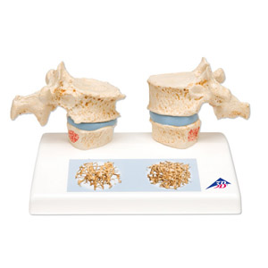
Impressive didactic model for comparing osteoporotic and normal thoracic vertebrae. Shows reproductions of osteoporotic 11th and 12th thoracic vertebrae with narrower intervertebrat disc located on the left of the stand.Two corresponding healthyvertebrae with intervertebral disc are provided on the right side. The upper vertebra is divided in the middle and can easiiy be opened thanks to magentic connections to show the cut surfaces to demonstrate sintering and osteophytes. A detailed illustration on the base depicts two 3D micro CT images obtained from biopsies of both a healthy and an osteoporotic bone.
14x9x10cm;d.2kg
|