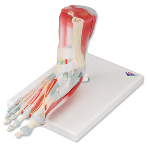
This anatomically detailed model of the foot and lower leg can be disassembled into 6 parts for detailed study of the following structures: bones, muscles, tendons, ligaments, nerves, arter-ies, and veins.
The frontal view of the foot model features the extensor muscles of the lower leg. The tendons can be followed on their passage under the transverse and crucial crural ligaments all the way to their insertion points. In addition all tendon sheaths of the foot area are visible.
On the dorsal portion of the foot the gastrocnemius muscle is removable to reveal deeper anatomical elements. The sole of the foot is represented in three layers:
・The first removable layer displays the flexor digitorum brevis
・The second removable layer consists of the quadratus plan-tae, the tendon of the flexor digitorum longus, and the flexor hallucis muscle
・The third layer reveals even deeper anatomical details of the foot
23 x 26 x 19 cm; 1.1 kg
|